
Virtual gel
The gel bench's plate as its own canvas: every lane on one migration scale, positioned by a real ladder, and honest about what the gel you described could actually resolve.
What it does
A virtual gel answers the question you would otherwise answer by running one: if I load these fragments on this gel, what will I see? Every band is a real fragment length — from a digest, a PCR, the open molecule, or a list you typed — placed by an electrophoretic mobility model rather than drawn where it looks convincing.
The plate is one shared coordinate space. The reference ladder and every sample lane migrate on the same scale, which is what makes a gel readable: you size a band by where it sits against the marks beside it, not by a number printed next to it.
When you'd use this
- Before you run the real gel. Choosing the % agarose is the whole game — a 1% gel will not separate a 180 bp fragment from a 220 bp one, and the plate shows you that before you cast it.
- To check a digest is diagnostic. Two constructs that differ by an inversion give different fragment sets; whether those sets are distinguishable on a gel is a separate question, and this is where you ask it.
- To read a result you already have. Load the lanes you ran and compare band positions against the same ladder your photograph used.
- As a figure. The plate is the picture that goes beside the protocol.
What this computes
- A migration distance per band, under one of two published mobility models — not two renderings of one: Helling–Goodman–Boyer, the classical log-linear relationship between fragment length and migration with a topology term for DNA form, or Rill–Van Winkle
- Ladder-calibrated positioning as the alternative to casting a gel: every band read off the ladder's own curve, in which case topology cannot move it — and the per-lane DNA form control says so rather than sitting there inert
- The resolving window of the % agarose you cast, reported on the status line, over 0.3–5.0 %
- Each band's size on hover, and the run description under the drawing naming the ladder that positioned it
- Per-lane DNA form — linear, supercoiled, nicked, relaxed — applied only where the model supports it
What it refuses, instead of drawing nothing: an empty bench draws no plate, and the canvas names the two ways to fill it.
Where it lives
It opens as its own canvas tab on the stage as soon as the bench has a lane, and the Virtual Gel Bench panel keeps every control. That split is deliberate: a plate gets wider with each lane added, and reading lane against lane is exactly what a 380 px side panel cannot do.
The tab and the panel are the same gel — there is one plate, not two. Change the ladder, the % agarose, the mobility law, a lane's DNA form or the render mode in the panel and the canvas follows immediately.
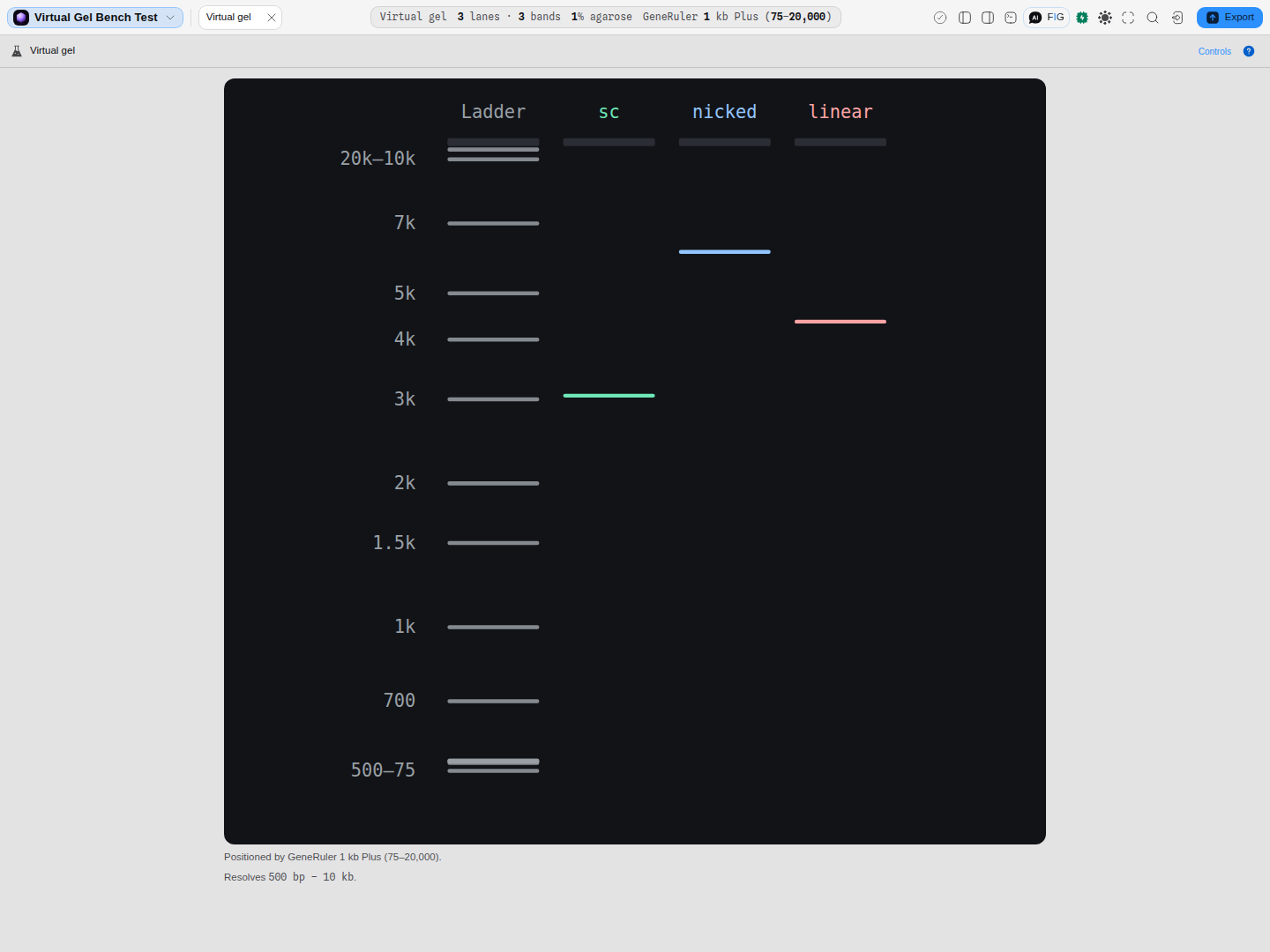
The same 4,361 bp circle in three lanes, at three different heights, with each lane named for the form it was loaded as. Topology is not size: the supercoiled lane is compact and runs ahead of the linear one, and the nicked circle drags behind both — so a single preparation can look like three different constructs on one gel, and reading the apparent size off the ladder would call them 3 kb, 4 kb and 6.5 kb. Per-lane form needs a cast gel to mean anything, which is why this one is at 1.0 % agarose: under ladder-calibrated migration every band is read off the ladder's own curve, so topology cannot move it, and the form control says so rather than sitting there inert. The plate scales to the tab, so the marks stay readable as lanes are added.
Inputs and outputs
In: lanes of fragment lengths. A lane can come from the open molecule (its whole length), a pasted list of bp values, a hand-off from a restriction digest or a PCR run, or your own typed marker.
Out: a positioned plate — every band's migration distance, its size when you hover it, and the run description under the drawing ("Positioned by GeneRuler 1 kb Plus").
What it refuses, and what it says instead of drawing nothing:
- An empty bench draws no plate. The canvas says so and names the two ways to fill it, rather than showing an empty rectangle that looks like a gel with nothing on it.
- A per-lane DNA form that cannot move anything is disabled with its reason on screen. Under the Rill–Van Winkle law the topology term drops out, so supercoiled, nicked and linear land on the identical migration distance; a live control there would be a knob that does nothing.
- A calibration table that cannot be used is refused next to the control that set it, never silently ignored.
Controls
| Control | What it changes |
|---|---|
| Ladder | Which of ten size standards provides the reference lane and the bp axis. |
| % agarose | The gel you are casting, 0.3–5.0%. This drives migration and the resolving window reported on the status line. Leave it unset to calibrate off the ladder's own marks instead. |
| Mobility law | Helling–Goodman–Boyer or Rill–Van Winkle — two published models, not two renderings. See below. |
| DNA form (per lane) | linear · supercoiled · nicked · relaxed. Topology changes effective mobility, which is why a supercoiled plasmid runs ahead of the same plasmid cut once. |
| Render mode | Bands (rectangles), raster (a photographic smear from the intensity profile), or both. The same three modes, in the same order, as the digest and PCR gels. |
| Brightness reference | Whole plate scales every band against the brightest band on the gel, so brightness stays comparable between lanes and a lightly loaded lane can draw almost black. Per lane scales each lane against its own brightest band, so every lane is legible and no between-lane comparison survives. The line under the plate names whichever is in force. |
| Calibrate | Supply your own (bp, Rf) table, from a gel you actually ran, and position every band against that instead of a catalogue ladder. |
| Add / rename / remove lane | The plate's contents. Lane order is the loading order. |
| Save / load gel | Named plates, so a run can be reopened rather than rebuilt. |
Algorithms you can choose
The two mobility laws answer the same question with different physics, and they disagree most at the extremes:
| Law | What it models | Where to prefer it |
|---|---|---|
| Helling–Goodman–Boyer | The classical log-linear relationship between fragment length and migration distance, with a topology term for DNA form | Routine analytical gels across the usual 0.5–2% range, and any run where supercoiled/nicked/linear must separate |
| Rill–Van Winkle | A reptation-based crossover model, valid over a narrower band of casting percentages | Runs where the classical law's log-linear assumption breaks down at the size extremes |
The topology term is real, and it is why the two laws differ visibly. The form factor multiplies effective molecular weight — supercoiled 0.7×, linear 1.0×, nicked and relaxed 1.4× — so under Helling–Goodman–Boyer one 4,361 bp plasmid draws at three distinct positions as supercoiled, nicked and linear. Under Rill–Van Winkle all three land on the same distance, and the workbench disables the per-lane control and tells you why rather than leaving a knob that moves nothing.
What the status line tells you
With a plate on screen the bar carries four readings, in the order you would check a gel:
- Gel — lanes and total bands: the shape of the run.
- Run — the % agarose and which law placed the bands, or ladder-calibrated when no % is in play.
- Positioned by — the ladder or your own calibration. Every bp you read off this gel is a claim about that reference, and this is where it is named.
- Outside the window — appears only when it applies. A gel at a given % resolves a finite range (1.5% agarose resolves roughly 200–4,000 bp); fragments outside it are still drawn, by the law's damped extrapolation, and they look exactly as trustworthy as the rest. This reading is what tells you they are not measurable — change the % agarose to bring them into range.
Pitfalls
- A band drawn is not a band you could measure. Extrapolated positions outside the resolving window are the most common way a virtual gel misleads. Watch the Outside the window reading.
- The ladder is the ruler, so changing it changes every size you read. The plate says which one positioned it for exactly this reason.
- Two fragments of similar size merge into one fat band, on a real gel and here. Raster mode shows that honestly; bands mode draws two rectangles that may overlap.
- A supercoiled plasmid runs "too fast" for its length. That is not an error — it is the form factor. If a band's apparent size disagrees with the construct, check the lane's DNA form first.
What it hands to and takes from other tools
- Restriction digest and PCR send their fragments here — the digest's fragment table and the amplicon set become lanes without retyping anything.
- The open molecule can be loaded as a lane at its full length, so a linearised backbone can be compared against its digest.
How we know it is right
Migration is computed from the published Rill / Van Winkle agarose mobility law (Electrophoresis 23(1) and 23(16), 2002), by the same model the digest and PCR gels use — so the three cannot disagree about where a fragment runs. The resolving windows are the published empirical ranges for each casting percentage, and the ladders are real catalogue products with their marks as the vendors publish them.
Digest gel
The restriction digest's own plate as a canvas: every lane you set up, cut on the molecule you have open, migrated against a real ladder at the % agarose you would cast.
Initiation-window fold
The mRNA secondary structure around a start codon, drawn as arcs on the sequence axis, so an occluded ribosome binding site is something you can see rather than infer from one energy.